
Chronic Back Pain Gold Coast —
For Complex & Persistent Cases
When your back presentation hasn’t responded to care — here’s a different approach. Assessment-led, movement-based, grounded in exercise science and remedial practice.
Most back pain responds well to standard care. But some cases are more complex. If yours hasn’t improved despite doing everything right, the assessment may need to go deeper than where it hurts. Based in Varsity Lakes, this practice supports clients across the Gold Coast.
What if the issue isn’t where it hurts?
The source of a movement limitation
isn’t always where symptoms are felt.
In this example, movement changed within the same session — even though the back wasn’t addressed directly. Ongoing assessment helped guide the approach as the body adapted.
This is the core of assessment-led practice: finding what’s contributing to the presentation, not just treating the site of discomfort.
Case study — de-identified, with written consent
From barely walking
to ultra trail running.
This case documents the progression of a 55-year-old recreational runner diagnosed with severe multi-level lumbar disc herniation. Prior treatment approaches had provided limited sustained improvement, and the clinical outlook at the time suggested a guarded prognosis regarding structural change.
Over a 39-month period of assessment-led management, the focus shifted toward restoring load tolerance, movement efficiency, and functional capacity. The progression below outlines the measurable observations that followed.
“At presentation she could barely walk and was in considerable distress about her capacity to return to running. Prior care had provided limited sustained improvement.”
— Adapted clinical record, November 2022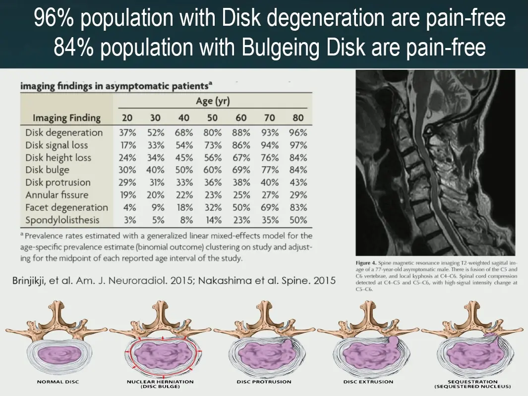
Understanding the research
Why back pain sometimes
doesn’t respond to standard care.
The answer is often not about the diagnosis on the imaging report. Population research (Brinjikji et al., 2015) consistently shows that disc bulges, herniations, and degenerative changes are found on MRI in people who have no discomfort at all:
Brinjikji et al. (2015), AJNR — peer-reviewed systematic review of imaging findings in asymptomatic populations.
This doesn’t mean imaging is useless — it means structural findings alone don’t explain why someone presents with discomfort, and treating the image rather than the person can miss the actual contributing factor.
Structural appearance does not define functional capacity. Imaging findings alone should not be used to limit what you believe is possible.
In complex or treatment-resistant cases, the real driver is often:
- Neuromuscular compensation patterns built up over years of guarding and load avoidance
- Fascial restrictions across the thoracolumbar chain, hips, and pelvis
- Load asymmetry — the body protecting one side by overloading the other
- Respiratory mechanics dysfunction affecting intra-abdominal pressure
- Speed- or load-dependent thresholds that only appear under demand
None of these are visible on imaging. All of them are identifiable through assessment.
The hip is a common example. The psoas, one of the main hip flexors, attaches directly to the lumbar spine, so tension at the front of the hip can change how the lower back moves when you walk or stand tall. An ache on one side of the lower back, just above the hip, is often reported alongside tight hip flexors. I look at the hip and lower back together — see hip flexor tightness and the lower back.
The assessment process
What a whole-body assessment
reveals in chronic back presentations.
Every first appointment begins with assessment before any hands-on treatment. For complex back presentations this includes:
- Medical history review and imaging analysis Understanding what’s been tried, what’s been found, and what may have been missed.
- Observational movement analysis Antalgic patterns, guarding, compensation strategies under load.
- Bilateral spinal palpation Tissue density, fascial restriction, and asymmetry across the lumbar and thoracic chain.
- Active and passive range of motion Throughout the lumbar, thoracic, and hip chain — not just the reported location.
- Respiratory assessment Diaphragmatic function and breathing pattern — often overlooked in back presentations.
- Functional movement screening Load avoidance patterns and motor compensation under demand.
- VALD ForceDecks bilateral force platform testing (where indicated) Objective measurement of force output asymmetry — identifying the side the body is protecting.
In the case documented above, this assessment identified a speed-dependent threshold (symptoms only above 8 km/h), eccentric deceleration variability (CoV 203.6% on force platform testing), respiratory dysfunction, and fascial restrictions across the sacroiliac joint, thoracolumbar fascia, and lateral chain.
“These factors are not always visible on imaging and may not be assessed in standard protocols.”
— Clinical observationClinical timeline
From barely walking
to 65 km ultra trail race.
This case unfolded across seven distinct phases over 30 months. The timeline below outlines the key milestones, clinical decisions, and measurable changes at each stage.
Pain-free walking
Returned to unrestricted daily movement without lumbar discomfort
16 km continuous run
Sustained 16 km running with minimal lumbar symptoms
Pain-free running
Progressive return to unrestricted recreational running
Marathon completed (42 km)
Full marathon completed without lumbar symptom limitation
65 km ultra trail completed
Stanley Monster Ultra — sustained performance at age 55
Structural Diagnosis
MRI Lumbar Spine (2017): severe multi-level disc herniation at L3/4 with neural canal compromise. Prognosis at the time indicated a guarded outlook regarding structural change.
Presentation
Presented with significant difficulty walking and low confidence in returning to running. Previous care had provided limited sustained improvement.
Objective Baseline
VALD ForceDecks bilateral squat testing revealed marked neuromuscular variability (eccentric deceleration RFD CoV 203.6%), indicating an inconsistent and unstable load absorption strategy. Right-dominant force asymmetry was 10–15%. Symptom threshold confirmed above 8 km/h running speed.
Functional Restoration
Running 14–16 km continuously (approximately 90 minutes) with minimal symptoms achieved within five months of commencing structured management.
Imaging Lag
CT Lumbar Spine (Feb 2024) continued to show degenerative changes. Functional capacity, however, had already improved significantly — demonstrating that imaging findings did not fully reflect clinical presentation.
High-Load Performance
VALD ForceDecks countermovement jump (CMJ) testing — a higher-demand plyometric task — demonstrated substantial improvement in neuromuscular control.
Eccentric deceleration variability (RFD CoV) reduced from 203.6% at baseline to 56%, indicating a markedly more consistent and stable load absorption strategy.
The client progressed to ultra trail running during this period. No symptom provocation occurred during plyometric testing or higher-load activities.
Peak Performance
42 km marathon completed without limiting lumbar symptoms.
65 km Stanley Monster ultra trail race completed.
Ongoing running participation without recurrence of limiting lumbar symptoms.
Objective measurement
What the VALD ForceDecks
assessment revealed.
Two VALD ForceDecks assessments across the treatment period provide before-and-after evidence of neuromuscular change. The follow-up assessment involved a significantly higher mechanical and neurological demand task — yet neuromuscular variability reduced markedly.
| Metric | Feb 2023 — Baseline | May 2024 — Follow-up |
|---|---|---|
| Test type | Bilateral squat (bodyweight) | Countermovement jump (higher demand) |
| Ecc. Decel. RFD CoV | 203.6% — highly inconsistent load absorption strategy | 56% — markedly improved consistency and control |
| Symptom status | Discomfort reproduced above 8 km/h running | No provocation on plyometric task |
This suggests improved load tolerance and movement control under stress — not simply short-term symptom reduction. In practical terms, the body demonstrated a more consistent and efficient strategy for absorbing and redistributing force, which aligned with the client’s ability to return to long-distance running without recurrence of limiting lumbar symptoms.
Objective measurement was used to guide clinical reasoning and monitor progression, rather than relying solely on subjective symptom reporting.
Treatment approach
What was addressed —
and how.
Treatment was directed at the neuromuscular compensation patterns, not the structural findings on imaging. The core components were:
- Myofascial release Thoracolumbar fascia, hip flexor complex, bilateral hamstrings, quadratus lumborum, and surrounding chains.
- Joint mobilisation — Grade I–II Lumbar and thoracic segments, sacroiliac joint, and hips.
- Respiratory retraining Diaphragmatic function restored to regulate intra-abdominal pressure and spinal loading.
- Load redistribution and movement retraining Rotational mobility, single-leg loading, oblique activation under load.
- Self-management tools Foam roller, S-ball techniques, and a graduated running progression.
Session frequency began fortnightly, progressively reducing to monthly then bi-monthly maintenance as functional capacity improved and sustained gains were demonstrated between sessions.
Is this right for you?
This approach may suit
people who:
Based in Varsity Lakes, this clinic supports clients from across the Gold Coast — including Robina, Burleigh Heads, Mudgeeraba, Palm Beach, and surrounding areas.
The first appointment is always an assessment before any hands-on treatment begins. Imaging is reviewed, history is taken, and the whole body’s movement is observed — then a session plan is built based on what the assessment actually finds.
Common questions
Back pain —
frequently asked.
Can a disc bulge improve without surgery?
Many disc changes seen on MRI are common in people without any discomfort. In some cases, functional capacity and load tolerance can improve significantly with movement retraining, load management, and progressive rehabilitation — even when structural changes remain on imaging.
Why does back discomfort return after treatment?
Sometimes the initial presentation settles but the underlying load asymmetry or compensation pattern hasn’t fully resolved. When training intensity or daily demand increases, symptoms can reappear. Assessment-led care looks for the pattern, not just the current episode.
Can I run with a lumbar disc herniation?
In some cases, yes — if load tolerance is rebuilt progressively and movement strategy is stable. A structured assessment helps determine readiness and identify what thresholds, if any, need to be addressed first.
Does imaging always explain back discomfort?
No. Structural findings such as disc bulges are common in people without symptoms. Imaging is one part of the picture, not the whole explanation. How the body moves under load is often more informative than what the scan shows at rest.
When should I see a practitioner about back discomfort on the Gold Coast?
If your presentation has persisted despite appropriate care, keeps returning, or limits training or daily movement — whether you’re based in Varsity Lakes, Robina, Burleigh Heads, Mudgeeraba, or elsewhere on the Gold Coast — a deeper assessment may help clarify what’s contributing.
How long does back discomfort usually last?
Back presentations vary significantly. Acute episodes often settle within weeks, but cases involving compensation patterns, load asymmetry, or chronic tissue changes can persist much longer without targeted assessment and a structured management approach.
Ready for a
fresh assessment?
If you’ve been searching for answers, the first appointment is always an assessment — reviewing your history, imaging, and how your whole body moves before any treatment begins.
Educational content only. Not medical advice. Individual results vary. The case study presented is de-identified and published with written client consent for clinical and educational purposes. Hill Yang is an ESSA Accredited Exercise Scientist (AES #17005) and Remedial Massage Therapist (MMA #031045). Always consult a qualified health professional for personal health concerns.